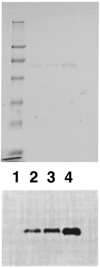
Figure 2.
SDS/PAGE analysis of low heparin and high heparin-affinity enzyme forms on a 12% polyacrylamide gel. Protein bands stained with Coomassie brilliant blue (Upper) and 75Se-labeled protein bands detected by PhosphorImager (Lower). Lane 1, prestained standard proteins (Novex) in 250, 98, 64, 50, 36, 30, 16, and 6 kDa; lane 2, 1 μg of high heparin-affinity HeLa cell enzyme (2,100 cpm); lane 3, 1 μg of low heparin-affinity human lung adenocarcinoma cell enzyme (1,200 cpm); lane 4, mixture of 1 μg of each (3,300 cpm).

