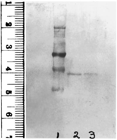
Figure 3.
Immunoblot analysis of low heparin-affinity enzyme forms. TR isolated from human lung adenocarcinoma cells and from HeLa cells was chromatographed on heparin agarose and 1 μg samples of enzyme fractions were subjected to electrophoresis and transferred to a poly(vinylidene difluoride) membrane followed by immunoblotting with a polyclonal anti-rat liver TR antibody (1:1,000 dilution). Goat anti-rabbit IgG (H+L), 5-bromo-4-chloro-3-indolyl phosphate, and nitroblue tetrazolium were used for visualization of reactive protein bands. Transferred standards: 98 kDa at 3.2 cm and 64 kDa at 3.9 cm (lane 1), low heparin-affinity enzyme from lung cells (lane 2) and from HeLa cells (lane 3). The high heparin-affinity enzyme forms were not detected.

