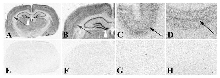
Figure 2.
Detection of miR-124a in rat and human brain sections using 33P-labeled RNA probes. A 21-nt RNA oligonucleotide complementary to miR-124a or a control 21-nt RNA oligonucleotide that contained three internal nucleotide mismatches to miR-124a were 5'-end labeled using T4 DNA kinase and γ33PATP. Approximately 1.0 × 10⁶ cpm of labeled probe were hybridized to adult brain sections from rat (miR-124a probe: A, B; mismatch probe: E, F) or human (miR-124a probe: C, D; mismatch probe: G, H) and processed for ISH as described in the text. Slides were air dried and exposed to BioMax MR film (Kodak) for 18 hours at room temperature prior to standard development. Images were collected on a flat bed scanner at 600 dpi. Prominent hybridization signals are present in distinct layers of cerebral cortex in both rat and human tissue sections with miR-124a probe (black arrows in A-D) but not with the miR-124a mismatch probe. Signal is also visible in distinct layers of the hippocampus. Note the intense labeling in the dentate gyrus (white arrowhead in A, B), with lower hybridization signals in other subfields of the hippocampus.

