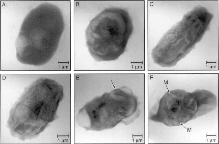
Figure 3.
Soft x-ray micrographs of P. falciparum malaria parasites infecting protein 4.1-deficient erythrocytes. (A) Two ring stage parasites infecting an elliptocytic erythrocyte. Structurally there are not obvious differences at the ring stage between parasites in normal and protein 4.1-deficient erythrocytes. Note the parasite apposed to erythrocyte membrane protuberance (exposure, 30 sec). (B–E) Trophozoites show structural derangements during maturation in protein 4.1-deficient erythrocytes. Parasites have a prolate spheroidal form (C and D) and complicated structures with redistributed mass not detected in trophozoites infecting normal erythrocytes. There is obvious damage to the erythrocyte membranes including (E), an image of a parasite that does not express major malarial proteins that associate with the erythrocyte membrane. Note the sharp edge (arrow) in E, demonstrating the resolving power of our microscope. The width of this edge is <80 nm (exposure: B, C, and E, 20 sec; D, 60 sec). (F) Multinucleated schizont in a protein 4.1-deficient erythrocyte. Arrangement of forming merozoites (M) and the residual body appears more disorganized than in normal erythrocytes (exposure, 30 sec).

