Figure 2.
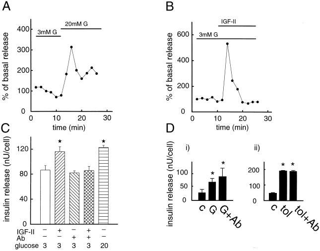
Effect of IGF-II on exocytosis of insulin. β cells were perifused in a buffer containing 3 mM glucose (G) for 10 min. The perifusion was continued in the presence of 3 (B) or 20 mM (A) glucose. Time of addition of IGF-II (50 ng/ml) (B) is indicated. Because glucose is the normal insulin secretagogue under physiological conditions, we used stimulation with a 20 mM concentration of the sugar as a positive control (A). Insulin release in A and B is expressed as percentage of basal secretion, defined as the average insulin release obtained at 3 mM glucose during the first 10 min of perifusion. Representative experiments out of three are shown. (C) Effect of IGF-II (50 ng/ml) on insulin release in cells that were preincubated in the absence or presence of an anti-IGF-II/M-6-P receptor antibody. Insulin is expressed as nanounits per cell. Insulin release was investigated in batch incubations. Mean values ± SD for one representative experiment in quadruples out of three are shown. (D) Influence of an anti-IGF-II/M-6-P receptor antibody on glucose-stimulated (20 mM) (i) and tolbutamide-stimulated (100 μM) insulin release (ii). The figure shows a representative experiment, out of three, in quadruples for glucose and tolbutamide, respectively. ∗, P < 0.05.

