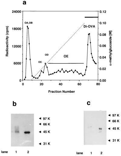
Figure 1.
Isolation and characterizations of Di-OVA isolated from hen oviduct. (a) Chromatogram of metabolically [3H]OVA on a Con A-Sepharose column. [3H]OVA was obtained by incubation of oviduct tissues with [2-3H]mannose at 37°C for 3 h according to Kato et al. (18) and was applied to a Con A-Sepharose column (0.9 × 15 cm), washed, and eluted with a linear gradient of α-methylglucoside from 0 to 0.1 M in 200 ml of 50 mM Tris⋅HCl (pH 7.2) containing 0.15 M NaCl, 0.1 mM CaCl2, 0.1 mM MnCl2, and 0.02% sodium azide, and then with 0.5 M α-methylmannoside in the same buffer. Fractions of 3 ml were collected and monitored by radioactivity. (b) SDS/PAGE of Di-OVA and OE as detected by fluorography. Lanes: 1, Di-OVA; 2, OE. Di-OVA was larger than OE by 2.0 kDa. Molecular weight markers are as follows: 97 kDa, phosphorylase B; 66 kDa, BSA; 45 kDa, OVA; 31 kDa, carbonic anhydrase. (c) Immunochemical identification of Di-OVA and OE with anti-OVA antiserum. Lanes: 1, Di-OVA; 2, OE.

