Figure 2.
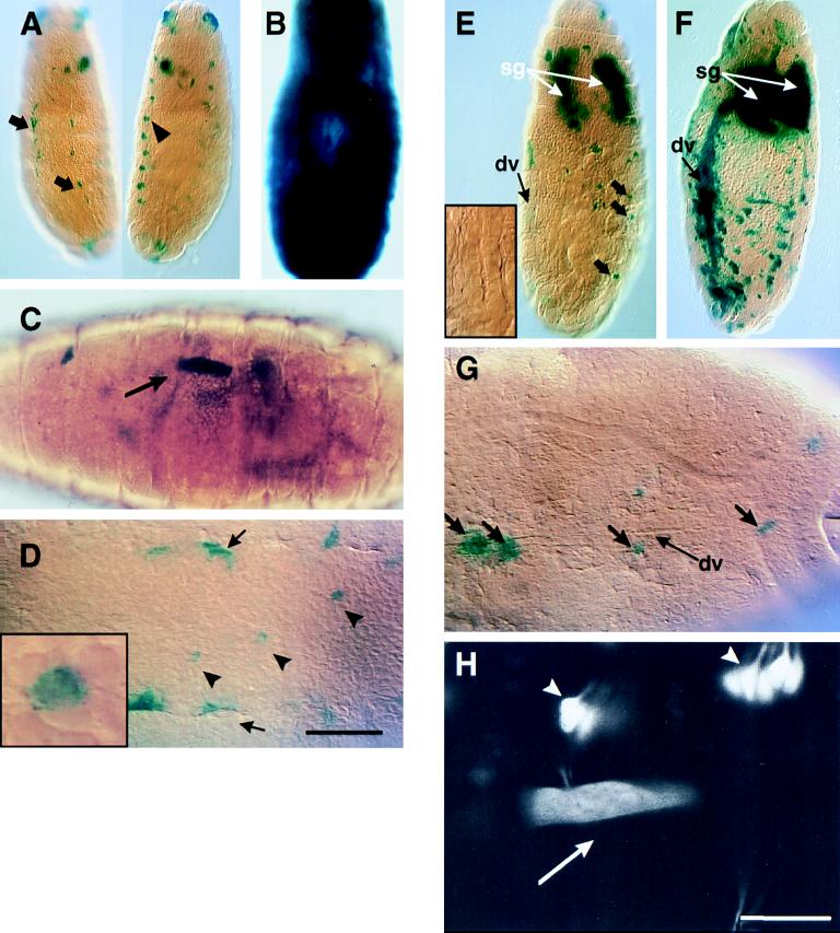
Induction of heat shock in either hsp26-lacZ embryos (A–D) or with the GAL4-UAS system (E–H). (A) Control embryos, maintained at 18°C without heat shock until fixation at stage 16. In the dorsal view (Right), non-heat-shock-dependent β-galactosidase expression can be seen in cells along the tracheal trunks (arrowhead) and in several dorsal anterior cells. The ventral view (Left) shows expression along the edges of the central nervous system (arrows), but not within it. (B) Late-stage embryo subjected to 20 min of standard heat shock. Cells throughout the embryo express β-galactosidase. (C) Induction of β-galactosidase expression in a single ventral somatic muscle fiber (arrow) by laser heat shock. Note that no other muscle fibers are stained. (D) Induction of β-galactosidase expression in a row of single cells within the central nervous system (arrowheads). The expression along the borders of the central nervous system (arrows) is non-heat-shock dependent. (Inset) A closeup of the center stained cell. The single-cell nature of the laser heat-shock induction can be seen clearly. (E) In the absence of heat shock, the GAL4 driver hsGAL4 N630 drives expression in a subset of sensory neurons (wide arrows) and in the salivary glands (sg, white arrows), measured here with a UAS-lacZ reporter gene. Note that there is no expression in the dorsal vessel and surrounding tissues (dv, black arrow; Inset). (F) When a 20-min heat shock is given, expression is induced most consistently in cells in the dorsal portion of the embryo, especially in and around the dorsal vessel (dv, black arrow), and more sporadically elsewhere throughout the embryo. White arrows indicate the salivary glands (sg). (G) Laser heat shock can induce expression of the UAS-lacZ target gene in specific single cells; here cells in the dorsal vessel (dv) were targeted (thick arrows). (H) Use of a UAS-GFP[S65T] target gene allows in vivo imaging of expression induced by laser heat shock. In this confocal micrograph, expression can be seen in a single ventral muscle fiber (muscle fiber 13, arrow). The non-heat-shock-dependent expression of the N630 driver is evident in the lateral chordotonal neurons and other sensory axons (arrowheads). (A–G) 5-bromo-4-chloro-3-indolyl-β-d-galactoside staining. (Bars: A, B, E, F = 100 μm; C = 60 μm; D = 30 μm; G = 35 μm; H = 25 μm.)

