Figure 2.
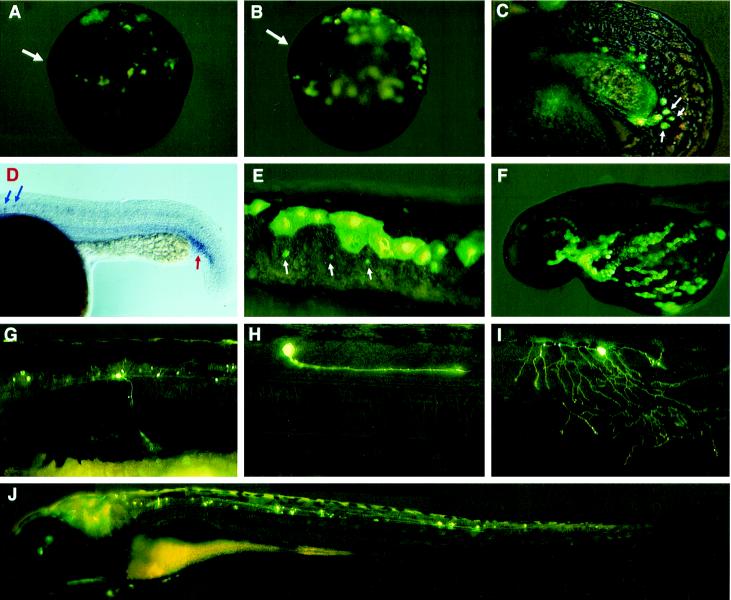
GFP expression driven from GATA-2 promoter constructs in living zebrafish embryos. (A and B) GFP expression driven by P1–GM2 in ventral ectoderm and mesoderm of dorsal shield stage embryos. Arrows indicate the dorsal shield. (C) GFP expression driven by P1–GM2 in hematopoietic progenitor cells located at the posterior end of ICM. (D) Expression pattern of GATA-2 in a 24-h embryo detected by RNA in situ hybridization. A red arrow indicates the ICM, and blue arrows indicate neuron cell bodies. (E) GFP expression driven by P1–GM2 in the EVL and neurons. Arrows indicate neuron cell bodies. (F) GFP expression driven by P3–GM2 in the EVL. (G–J) GFP expression driven by nsP5–GM2 in different types of neurons. In most injected embryos, fluorescent secondary motoneurons were observed (G). Occasionally, a fluorescent descending interneuron (H), Rohon–Beard cell (I), or neuron extending from the brain into the trunk (J) was observed. Neuron clusters were also observed in the brain and eyes.

