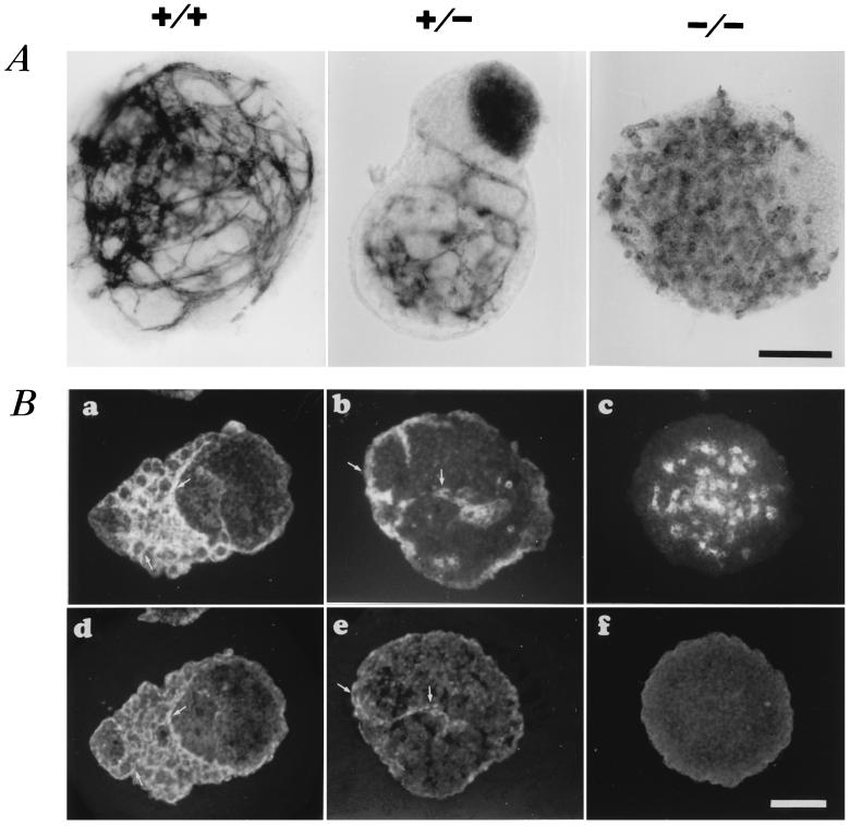
Figure 2.
Effect of VE–cadherin null-mutation on vascular phenotype in ES cell-derived, 11-day-old EBs. (A) PECAM whole mount immunocytochemistry of representative wild-type+/+, VE–cadherin mutant+/−, and −/− ES-derived, 11-day-old EBs. (B) CD34 (a–c) and VE–cadherin (d–f) immunofluorescence expression pattern in VE–cadherin+/+, +/−, and −/− EBs on serial sections. Arrows point to cord-like structures that coexpress both markers. No detectable staining with VE–cadherin antibodies can be observed in homozygous VE–cadherin−/− EBs. (Bar = 100 μm.)

