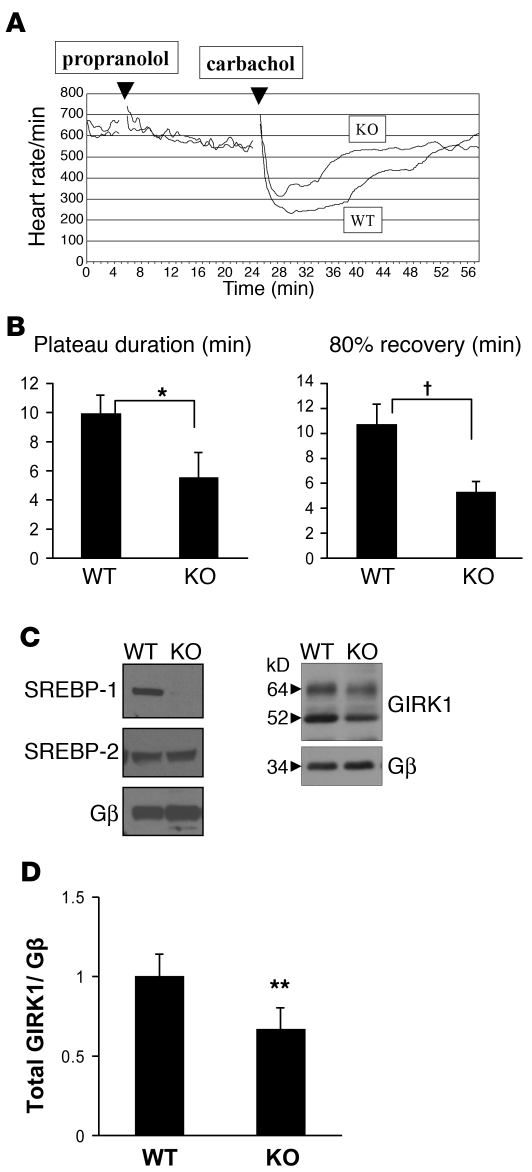
Figure 5. Response of the heart to parasympathetic stimulation and the expression of GIRK1 in SREBP-1 KO mice.
(A) Comparison of the negative chronotropic response of WT and SREBP-1 KO mice to carbamylcholine. Continuous ECGs were recorded as described in Methods. Mice were treated with an intraperitoneal injection of propranolol, 1 mg/kg, and 20 minutes later by intraperitoneal injection of carbachol, 0.2 mg/kg. Data represent the mean heart rate of 7 age-matched mice. (B) Quantitation of the negative chronotropic response to carbachol in WT and SREBP-1 KO mice. The plateau time of bradycardia and the time elapsed for 80% (Rec80) recovery to baseline heart rate, defined as heart rate following propranolol treatment, were determined as described in Methods. *P < 0.05; †P < 0.05. (C) Expression of SREBP-1 and GIRK1 in atria of WT and SREBP-1 KO mice. Atria of age-matched male WT and SREBP-1 KO mice were harvested and homogenized, and SREBP-1 (left panel) and GIRK1 (right panel) were determined by Western blot analysis. (D) Mean intensity of both glycosylated and unglycosylated GIRK1 bands determined by densitometry analysis of autoradiographs from 8 WT and 8 SREBP-1 KO mice normalized to the expression of Gβ; **P < 0.03.

