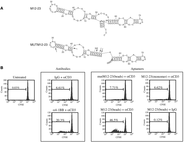
Figure 2. Characterization of M12-23 and mutM12-23 aptamers.
(A) Sequence and computer-predicted secondary structure. Arrows show the nucleotide changes introduced into the M12-23 sequence to generate mutM12-23. (B) CFSE proliferation assay of suboptimally activated CD8+ T cells incubated with bead-multimerized 4-1BB aptamers. CD8+ T cells isolated from the spleens and lymph nodes of Balb/c mice were labeled with CFSE and cultured in the absence or presence of either 1 μg/ml anti-CD3 or isotype-matched hamster IgG control Ab. At 16 hours after plating, the following reagents were added, as indicated: 5 μg/ml anti–4-1BB Ab or isotype-matched rat IgG2a Ab, or 100 nM M12-23 aptamer (monomer), bead-coupled M12-23 aptamer, or bead-coupled mutM12-23 aptamer. Cellular fluorescence was measured with flow cytometry 72 hours after plating. Percentages within each panel indicate the fraction of cells that underwent proliferation. See Methods for additional details.

