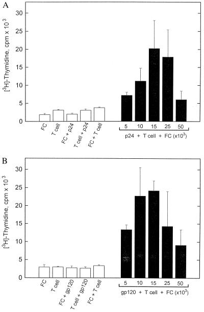
Figure 4.
Murine fibrocyte antigen presentation in vitro. Murine T cells (2 × 10⁵) purified from the spleens of p24 (A) or gp120 (B) immunized BALB/c were incubated with 2 μg/ml p24 or gp120 together with various numbers of mitomycin C-treated autologous fibrocytes. After incubation for 5 days, the cultures were pulsed for 12 hr with 4 μCi/ml [3H]thymidine and cell proliferation analyzed by liquid scintillation counting. Controls are illustrated on the left side of each figure. Data are expressed as mean ± SD and are representative of one experiment that was performed three times.

