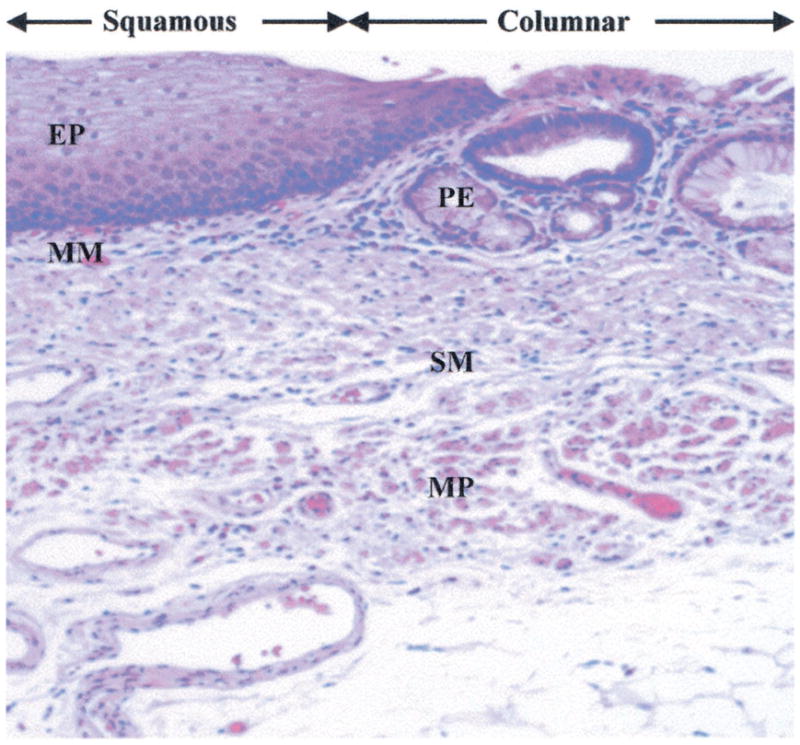
Fig. 6.

Histology showing squamous epithelium (EP) and muscularis mucosa (MM) on the left, columnar mucosa with pit epithelium (PE) on the right, and submucosa (SM) and muscularis propria (MP) on both sides.

Histology showing squamous epithelium (EP) and muscularis mucosa (MM) on the left, columnar mucosa with pit epithelium (PE) on the right, and submucosa (SM) and muscularis propria (MP) on both sides.