Figure 1.
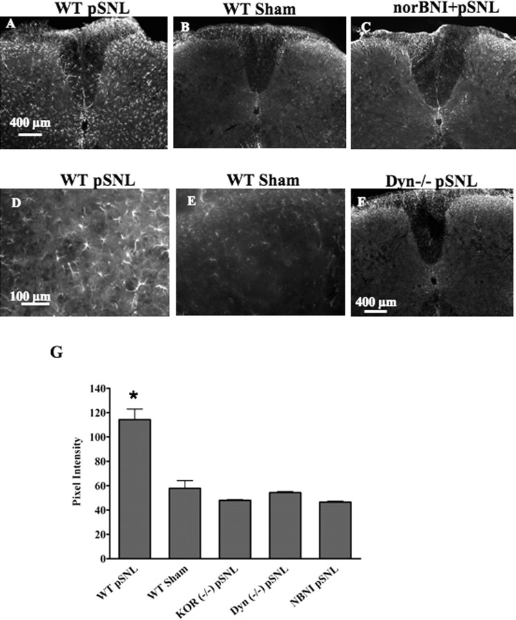
GFAP-immunoreactive staining is increased in WT lumbar spinal cord 7 d after pSNL but not after sham ligation. The increase is blocked by norBNI and does not occur in Dyn−/− or KOR−/− mice. Transverse sections were taken of the lumbar spinal cord 7 d after pSNL. A–C,GFAP staining was significantly increased in WT pSNL mice compared with WT sham-operated mice (A, B), and this increase in GFAP-IR was blocked by the κ antagonist norBNI (10 mg/kg) (C). Mice lacking the dynorphin gene (Dyn−/−) did not show upregulated GFAP staining after pSNL (F). D and E are higher-magnification images of A and B, respectively. G, Graph showing mean ± SEM pixel intensity of GFAP staining in lamina I–IV of lumbar spinal sections. Image analysis of astrocytes expressing GFAP after pSNL, demonstrating approximately twofold higher signal intensity [114 ± 8.71 arbitrary units (AU); n = 4] than that of sham-ligated mice (57.9 ± 6.26 AU; n = 4) (*p < 0.001). In contrast, image analysis of sections from pSNL mice pretreated with norBNI (NBNI) did not show a significant increase over sham and were significantly lower than WT pSNL signal intensity (norBNI, 46.5 ± 0.85 AU; n = 4). Similarly, pSNL of Dyn−/− or KOR−/− mice did not result in significant increases in GFAP-immunoreactive pixel intensities over sham-ligated mice (Dyn−/−, 54.3 ± 0.84 AU, n = 4; KOR−/−, 48.0 ± 0.62 AU, n = 4). Scale bars: A–C, F, 400 μm; D, E, 100 μm.

