


FIG 2.
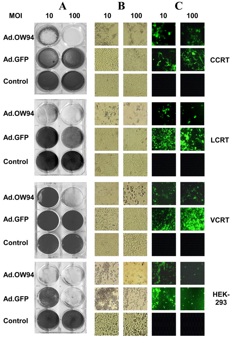
Lytic activity of Ad.OW94 (E1+) or Ad.GFP (E1−) on CCRT, LCRT, VCRT, and HEK-293 cells. Each of the cell lines were seeded at 2 × 10⁵ cells per well onto 6 well plates and infected with Ad.GFP or Ad.OW94 at MOI of 10 or 100 PFU per cell, or treated with a PBS control. After 7 days, cells were: (A) stained with 1% crystal violet, (B) examined microscopically for CPE, and (C) examined by fluorescence microscopy for GFP expression.