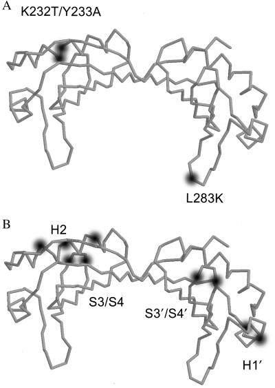
Figure 1.
Structure of TBP indicating the position of the mutations. The diagram was obtained with the program rasmol (52) with coordinates of the yeast TBP (53). For a comparison of the numbering of the residues of yeast and human TBP see ref. 54. The mutations H2, H1′, S3/S4, and S3′/S4′ are alanine-scan mutations described by Tansey et al. (36). H2 is a triple mutation (R231A, R235A, and R239A). H1′ is a double mutation (R269A and E271A). S3/S4 is a double mutant (E206A and R208A). S3′/S4′ is a double mutant (K297A and R299A). L283K is a mutation equivalent to the yeast mutation L189K (34) and K232T/Y233A is equivalent to the yeast mutation N2–1 (35). The use of the mutant L283K is based on the assumption that it is function for basal transcription as has been demonstrated for the yeast counterpart.

