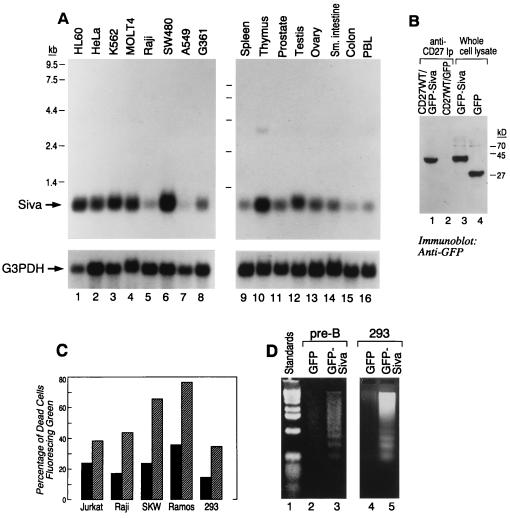
Figure 3.
(A) mRNA expression of Siva by Northern blot analysis is shown (upper bands) and is compared with the expression of G3PDH (lower bands). (B) CD27 binds to Siva in 293 cells. CD27WT DNA was transiently coexpressed with either GFP or GFP–Siva for 2 days. The CD27 receptor expression appeared to be similar as seen by flow cytometric analysis (data not shown). CD27 immunoprecipitates using anti-CD27 antibody (IA4) were separated by SDS/PAGE, transferred, and blotted with anti-GFP antiserum. Similar amounts of GFP and GFP–Siva were expressed as seen in the lanes corresponding to whole-cell lysates (lanes 4 and 3). CD27 coprecipitaed GFP–Siva (lane 1) but not GFP (lane 2). (C) Siva-induced apoptosis. Jurkat, Raji, SKW, Ramos, and 293 cells were transiently transfected (2 days) with GFP (hatched bar) and GFP–Siva (solid bar). The histograms represent the percentage of dead cells (based on cell morphology) in the cells expressing green fluorescence. In the case of 293 cells, along with GFP and GFP–Siva, pSVb containing the β-galactosidase gene was cotransfected. β-Galactosidase expression was visualized by X-Gal. The percentage of rounded shrunken cells in the fluorescent population of cells is presented. (D) Transient expression of GFP–Siva but not GFP can induce apoptosis in 293 cells (lane 3 vs. lane 2), the murine pre-B cell line (lane 6 vs. lane 5), and Raji cells (lane 8 vs. lane 7), as judged by the generation of DNA ladder.

