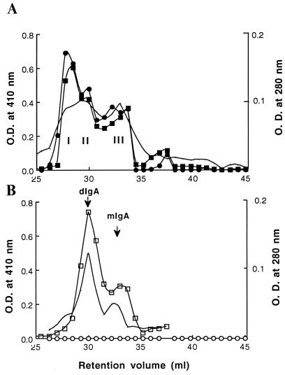
Figure 2.
Analysis of the composition of proteins secreted by transfectants synthesizing chimeric sIgA1 (A) or IgA1 (B). Three hundred microliters of 100-fold concentrated serum-free medium was separated on the basis of molecular weight on two Pharmacia Superose 6 columns in series. The lines with no symbols indicate the elution profiles at 280 nm. Fractions of 0.75 ml were collected and analyzed by ELISA, and IgA was captured on dansylated-BSA-coated microtiter plates that were detected with rabbit anti-κ (squares) (Sigma) or anti-SC (circles), followed by goat anti-rabbit antibody conjugated to alkaline-phosphatase and substrate. The resulting O.D. at 410 nm is plotted. Closed symbols indicate sIgA and open symbols indicate IgA1. The presence of dIgA and mIgA in the peaks was confirmed by analysis of the fractions by SDS/PAGE.

