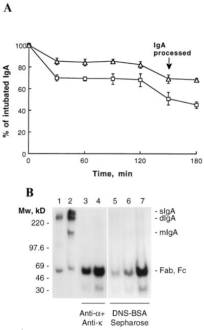
Figure 4.
In vivo stability of sIgA. (A) 125I-labeled dIgA (squares) and sIgA (triangles) were introduced directly into the stomach of BALB/c mice by intubation. IgA remaining in the mice was determined by whole body counting. Data are expressed as mean ± SD (n = 3). (B) After 150 min, a mouse intubated with dIgA (lanes 3 and 6) and a mouse intubated with sIgA (lanes 4 and 7) were killed and the intestinal washings isolated and processed as described. IgA from the intestinal washes was immunoprecipitated with either anti-α and anti-κ antibodies (lanes 3 and 4) or with DNS-BSA-Sepharose (lanes 6 and 7). For comparison, mice injected i.v. with radiolabeled dIgA were sacrificed after 3 hr, and the antigen-specific IgA was precipitated from the intestinal washes as above (lane 5). Half the precipitated proteins were analyzed by SDS/PAGE in phosphate gels. The gels were dried and exposed to Amersham Hyperfilm-MP for 48 hr. Also shown are the iodinated dIgA (lane 1) and sIgA (lane 2) used for intubate. The prestained molecular mass protein standards (Amersham) are indicated on the left; the positions of sIgA, dIgA, mIgA, Fab, and Fc, on the right.

