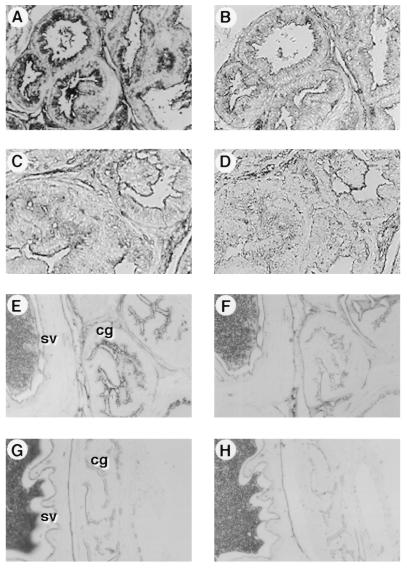
Figure 4.
Localization of PSA expression in the prostate and coagulating gland of the PSA1 transgenics by immunohistochemical staining. Formalin-fixed, paraffin-embedded tissue sections from the prostate (A and B) and coagulating gland/seminal vesicle (E and F) of a P1–9 transgenic, and prostate (C and D) and coagulating gland/seminal vesicle (G and H) of a nontransgenic control were incubated with rabbit anti-human PSA (A, C, E, and G) or control normal rabbit immunoglobulin (B, D, F, and H) followed by HRP-conjugated goat anti-rabbit Ig. Staining was visualized by adding the chromogen diaminobenzidine (A–D) or 3-amino-9-ethylcarbazole (E–H). (E and G) cg, coagulating gland; sv, seminal vesicle.

