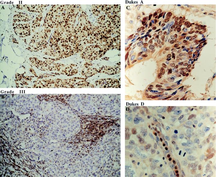
Figure 5.
Immunohistochemistry staining of p27kip1 in human breast and colorectal tumors using anti-p27kip1 mAb SX53G8. (A) Strong nuclear staining of p27kip1 in majority of the tumor cells in grade II invasive ductal carcinoma. (B) In grade III invasive ductal carcinoma, tumor cells are negative for p27kip1, while stromal lymphocytes are strongly stained. (C and D) Nuclear p27kip1 expression is also seen in Dukes A but not Dukes D human colorectal tumors. (A and B, ×250; C and D, ×600.)

