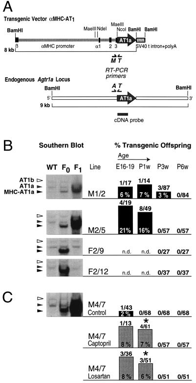
Figure 1.
Transmission rate and survival of αMHC-AT1 transgenic mice. (A) Transgenic vector. The complete intergenic region between the β- and α-myosin heavy chain genes (αMHC promoter) was used as a cardiac-specific promoter to control expression of the mouse AT1a angiotensin receptor. At the 3′ end of the AT1a receptor cDNA, the intron and polyadenylylation signal of the SV40 T antigen was added (SV40 t intron+polyA). β, α1,2,3 exons of the βMHC and αMHC locus; Agtr1a, angiotensin AT1 receptor gene; A, T, M, primers for reverse transcriptase–PCR. (B and C) Transmission frequency of the αMHC-AT1 transgene. Transgenic founder mice were mated with wild-type FVB/N mice, and the frequency of transmission of the transgene to their offspring was determined by Southern blot analysis using part of the coding region of the AT1a receptor cDNA. The endogenous AT1a gene can be detected as a 9-kb BamHI fragment (shaded arrowhead), the AT1b gene appears as a faint band at 10.6 kb size (open arrowhead), and the αMHC-AT1 transgene is detectable as a 8-kb fragment (solid arrowhead). For lines F2/9 and F2/12, blots from the F1 generation represent nontransgenic offspring, as no transgenic mice were born from these lines. Fifteen micrograms of genomic DNA was loaded per lane. Transmission rates are given in absolute numbers (number of transgenic offspring/total mice screened) and in percent values for the following age groups: at embryonic days 16–19 (E16–19), at 1 week after birth (P1w), at weaning age (3 weeks, P3w), and at 6 weeks of age (P6w). (C) Effect of ACE inhibitor treatment or AT1 receptor blockade on the survival of transgenic offspring of the M4/7 line. Pregnant mice were treated from day 12 after conception with captopril, losartan, or saline throughout pregnancy and during the lactation period. With both captopril and losartan, survival of the transgenic mice at 1 week of age was significantly improved as compared with untreated mice (∗, P < 0.05).

