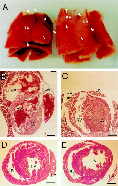
Figure 3.
Cardiac phenotype of the αMHC-AT1 transgenic mice. (A) Heart and lung preparation of transgenic (Left) and nontransgenic (Right) littermates from line M4/7 at day 4 after birth. The mother of these mice was treated with captopril from day 12 of pregnancy until day 4 after delivery (1 mg/ml drinking water). Compared with the control specimen, the transgenic heart displays a massive enlargement of left and right atria. Arrowheads mark the borders of the atria. RA, right atria; LA, left atria; V, ventricle. Bar = 2 mm. (B and C) Frontal section through the same transgenic (B) and nontransgenic (C) hearts as shown in Fig. 3A. The atrial cavity is greatly increased in transgenic atria compared with nontransgenic atria. RA, right atria; LA, left atria; LV, left ventricle; RV, right ventricle. Bar = 1.5 mm. (D and E) Horizontal cross-sections through the ventricles of transgenic (D) and nontransgenic (E) offspring from line M4/7 do not reveal any significant morphological differences between transgenic and nontransgenic ventricles. Bar = 1.2 mm.

