Figure 4.
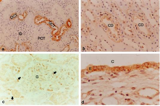
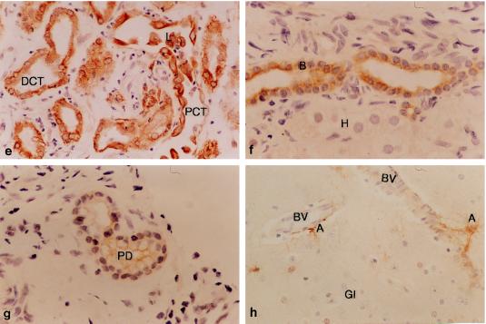
Polycystin expression in normal human and ADPKD tissues. All sections were formalin fixed and pretreated with trypsin as described, except c, which was fresh tissue. All sections shown are stained with antibody anti-FP-LRR except c, which is stained with anti-FP-46-1c. (a) Normal adult kidney showing staining of DCT. PCT and glomeruli (G) are negative. (b) Higher power view of normal CD demonstrating clear membrane staining. All cell surfaces are positive. (c) A glomerulus (G) from a fresh tissue specimen demonstrates staining of peritubular and glomerular endothelial cells (arrowed). (d) Section of renal cyst from end-stage ADPKD showing staining of cyst-lining epithelial cells (E). C, -cyst lumen. Cytoplasmic and membrane staining is still apparent but there is absence of staining in some cysts (data not shown). (e) Polycystin expression in acute tubular necrosis showing staining of DCT and regenerating and necrotic cells from the PCT. Necrotic cells can be seen in the PCT lumen. (f) Staining in normal liver is confined to the biliary epithelium (B) and not seen in hepatocytes (H). (g) Membrane staining is also seen in pancreatic duct (PD) epithelial cells and not glandular tissue. (h) Astrocytes (A) stain positively for polycystin with more intense staining seen in the perivascular cells. No neuronal or glial cell staining is seen (Gl, glial; BV, blood vessel).


