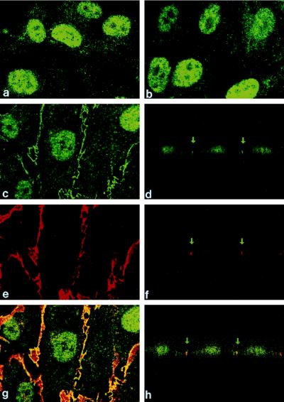
Figure 5.
Polycystin expression in cultured HUVEC. All figures demonstrate polycystin expression in HUVEC using anti-MAL-BD3 antibody. (a) HUVEC stained with secondary antibody alone demonstrating the nonspecific pattern of nuclear and cytoplasmic staining. (b and c) HUVEC stained with anti-MAL-BD3 antibody with or without preabsorption with fusion protein MAL-BD3 demonstrating the specific punctate linear expression pattern of polycystin. (e and g) Staining with PECAM (red) a lateral membrane marker colocalizes polycystin to the lateral membrane using two-color fluorescence (yellow). (d, f, and h) Vertical sections of c, e, and g also show the colocalization of polycystin and PECAM to the lateral membranes with no apical or basal staining.

