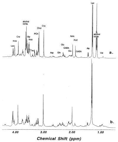
Figure 2.
Proton MR spectra (400 MHz) of human brain tissue collected from superior temporal gyrus histologically determined to be mildly affected with Pick disease. Spectrum a, intact tissue T2-weighted HRMAS at 2.5 kHz, at 2°C; spectrum b, tissue extraction solution at 20°C. Selected metabolite resonances were labeled on spectrum a (Lac:, lactate; GABA: γ-aminobutyric acid; Acet, acetate; Cre, creatine; Chol, choline; PCh, phosphorylcholine; Tau, taurine; Inos, inositol; alcohol conta: alcohol contamination). The resonances at 1.18 ppm (triplet) and 3.65 ppm (quartet) were only seen in the tissue spectrum, and were due to contamination with small amount of alcohol sprayed on the surface of the postmortem brain before tissue freezing (9).

