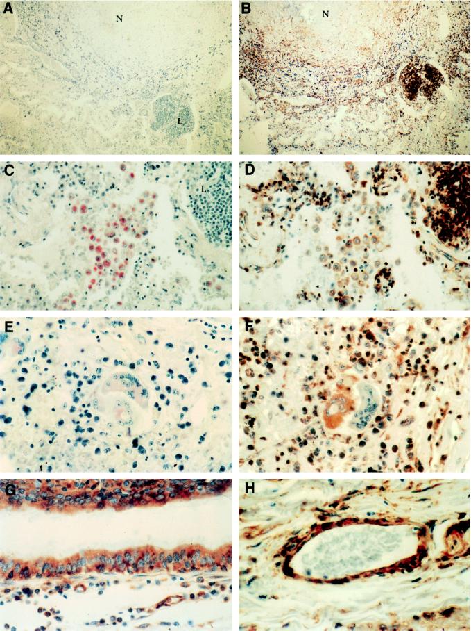
Figure 4.
Immunohistochemical stain of human tuberculosis specimen. (A) Tuberculosis granuloma stained with isotype control antibody showing hematoxylin counterstaining of nuclei, ×90. Area of caseating necrosis (N), lymphoid aggregate (L). (B) Serial section of granuloma in A, stained for osteopontin with mAb MPIIIB101, ×90. (C) Serial section of same specimen stained with anti-CD68 first-step antibody demonstrating macrophage aggregate, ×360. (D) Higher power view of B showing same macrophage aggregate staining for osteopontin, ×360. (E) Giant cells in specimen stained for CD68, ×900. (F) Higher power view of B showing osteopontin staining of epithelioid, but not Langhans, giant cell, ×900. (G) Small airway respiratory epithelium stained for osteopontin; same section though different field of B, ×900. (H) Small vessel staining with MPIIIB101, ×900. Similar results were obtained with sections from multiple specimens and patients.

