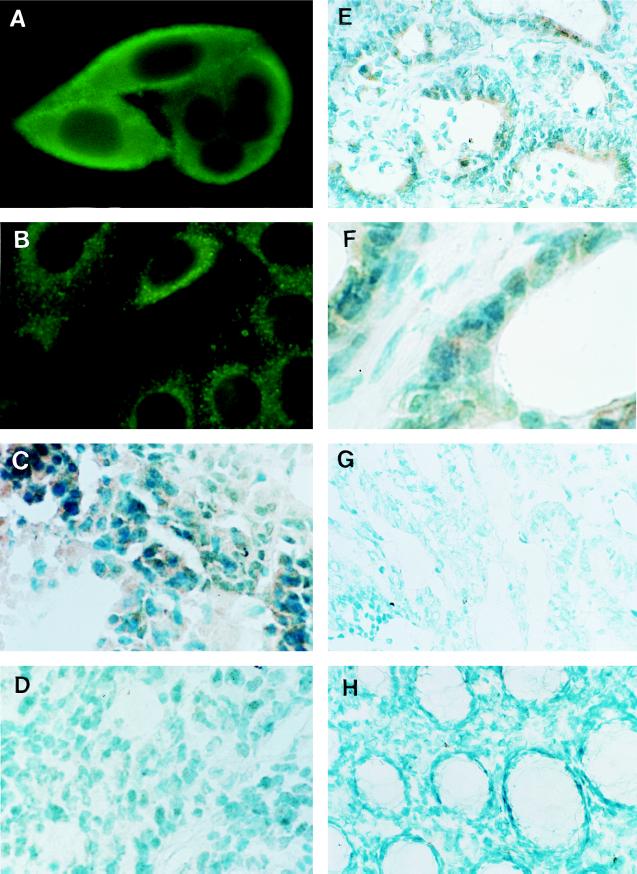
Figure 3.
Expression of FasL protein in human colonic adenocarcinoma. (A) Immunofluorescence staining of live SW480 human colonic adenocarcinoma cells with anti-FasL C-20 polyclonal antibody (×1,000). (B) Immunofluorescence staining of permeabilized SW480 cells with anti-FasL N-20 polyclonal antibody (×1,000). (C and D) Immunoperoxidase staining of cryostat sections of SW480 (C) and KM12C (D) xenograft tumors with anti-FasL N-20 polyclonal antibody (×400). (E and F) Immunoperoxidase staining of hepatic metastatic lesion of human colonic adenocarcinoma tissue (case 4 in Fig. 2) with anti-FasL N-20 polyclonal antibody (×200). F is a higher magnification (×400) of E. Note that especially as seen in F, every tumor cell strongly expressed FasL protein. (G and H) Immunoperoxidase staining of primary tumor of human colonic adenocarcinoma tissue (G) and normal human colon mucosa (H) with anti-FasL N-20 polyclonal antibody (×200).

