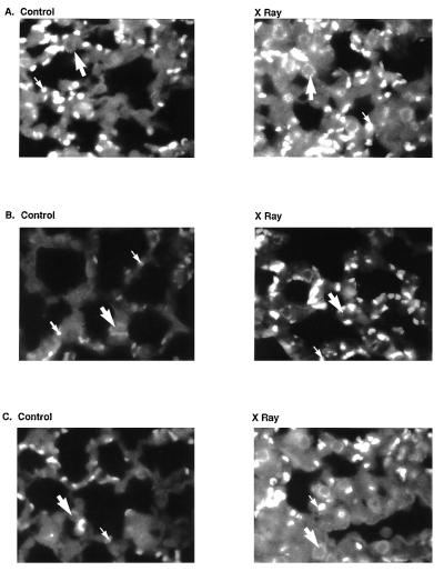
Figure 6.
Immunofluorescent staining for inflammatory cell infiltration by use of anti-CD45 in nonirradiated control and irradiated lungs from (A) ICAM+/+ mice, (B) ICAM−/− mice, and (C) ICAM+/− mice. Five weeks after treatment, lungs were fixed, sectioned, and stained with anti-CD45 antibody using rhodamine conjugates. Shown are photomicrographs of UV microscopy. Large arrows indicate CD45 on inflammatory cells, and small arrows indicate erythrocytes which demonstrate autofluorescence.

