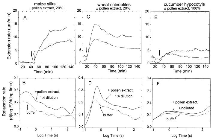
Figure 2.
Enhancement of cell wall extension (A, C, and E) and stress relaxation (B, D, and F) by maize pollen extract. A and B show rheology responses of maize silk walls to pollen extract diluted to 20% strength (1:4 dilution with 50 mM acetate buffer, pH 4.5). C and D show responses of wheat coleoptile walls to 20% pollen extract. E and F show the modest responses of cucumber hypocotyl walls to undiluted (100%) pollen extract. For the extension assays, heat-inactivated wall specimens were clamped in a constant-load extensometer in 50 mM sodium acetate buffer, pH 4.5; wall extension (creep) was detected by a position transducer attached to one of the clamps and is plotted as extension rate (9, 15). At the time indicated by the arrow, the buffer surrounding the wall specimen was exchanged for a similar one containing maize pollen extract. Extension traces show two representative results from four to eight replicates. For the stress-relaxation assays, heat-inactivated walls were preincubated in buffer ± pollen extract and then clamped in an extensometer, extended to a predetermined load, and held at constant length during the subsequent relaxation (15) either in 50 mM acetate buffer (dotted lines) or the same buffer containing maize pollen extract at the dilution indicated. The decay in stress is plotted as a relaxation spectrum (log-time derivative of stress). Each relaxation curve is the average of six to nine independent relaxation measurements.

