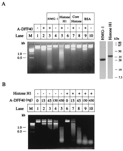
Figure 3.
Stimulation of DNase of DFF40 by histone H1 and HMG-1. (A) An aliquot (2 μg) of plasmid DNA (pcDNA3 vector) was incubated with either a 45-ng aliquot of A-DFF40 purified through the Mono S column as described in Fig. 2 in 30 μl of buffer A at 37°C for 2 hr alone (lane 2) or in the presence of 1 μg of bovine HMG-1 (lane 3), 0.5 μg of bovine histone H1 (lane 5), 1 μg of core histone (lane 7), or 1 μg of BSA (lane 9). The plasmid DNA was also incubated with buffer alone (lane 1), 1 μg of bovine HMG-1 (lane 4), 0.5 μg of histone H1 (lane 6), 1 μg of core histone, or 1 μg of BSA (lane 10). The reactions were stopped by adding 5 mM EDTA and the products were directly loading onto a 1.2% agarose gel containing 5 μg/ml ethidium bromide. An aliquot (5 μg) of bovine HMG-1 or histone H1 was also directly subjected to SDS/PAGE followed by Coomassie brilliant blue staining (the right two lanes with molecular mass markers). (B) An aliquot (2 μg) of plasmid DNA was incubated with the indicated amount of A-DFF40 purified through the Mono S column as described in the legend Fig. 2 in the absence (lanes 2–5), or presence (lanes 6–10) of 0.5 μg of histone H1 in a final volume of 30 μl of buffer A at 37°C for 2 hr. The reactions were stopped by adding 5 mM EDTA and the products were directly loaded onto a 1.2% agarose gel containing 5 μg/ml ethidium bromide.

