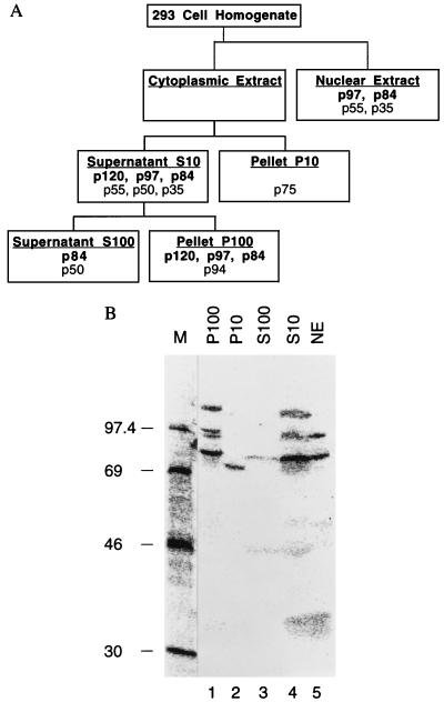
Figure 1.
Detection and subcellular localization of VA RNAII binding proteins. (A) Fractionation scheme showing the distribution of VA RNAII binding proteins. (B) Autoradiogram of a Northwestern blot, containing proteins from the indicated 293 cell fractions, probed with 32P-labeled VA RNAII. Lane M contains marker proteins whose molecular masses are indicated in kDa.

