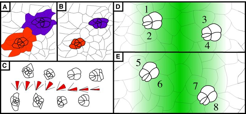
Figure 4. The interface for rotation resides between the photoreceptors/cone cells and undifferentiated cells.

(A) and (B): Two models illustrating potential interfaces between rotating cells. In (A) like-colored cells are linked together and move as a group so the interface is at the purple/orange border. In (B), undifferentiated cells do not rotate with differentiated cells. The interface between rotating and stationary cells is between the colored and uncolored cells. (C) Average angle of orientation by developmental stage. Each “stick diagram” corresponds to the ommatidial form that lies directly above or below the diagram. In the diagrams, the center line indicates the average angle, red shading highlights standard deviation, and outermost lines indicate minimum (lower line) and maximum (higher line) degree of rotation seen for each developmental stage indicated. (D) and (E): Schematic representation of relative likelihood of anterior and posterior cone cells being recruited from the BUdR-labeled pool of cells. Green-colored gradient represents probability of a cell being labeled with BUdR. In both panels, two ommatidial precursors are shown, one anterior to and the second posterior to, the BUdR-labeled band. Darker cells are more likely to be labeled since they lie closer to this band. Numbers identify cells that will become anterior and posterior cone cells. In (D), cells are recruited prior to rotation (as modeled in Fig. 4B), so cells numbered 1 through 4, which have equivalent levels of shading, share equal probabilities of being labeled. When examined as pupal eyes, equal numbers of labeled anterior and posterior cells are predicted. In (E), cells are recruited after rotation begins (as modeled in Fig. 4C). Consequently, two cells are recruited from the pool that is more likely to be labeled (6,7, dark green) whereas the other two cells are recruited from the pool that is less likely to be labeled (5,8, pale green). When examined as pupal eyes, the number of labeled anterior and posterior cells is predicted to be disproportionate. See text for details. Anterior is to the right for all panels.
