Figure 5.
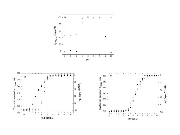
pH-, GdnHCl- and urea- induced unfolding of dimeric and tetrameric PfGST at pH 8.0 and 25°C. A. Effects of the pH on the CD signal at 222 nm for the dimeric (solidsquares) and tetrameric (hollow squares) PfGST. The data is presented as percentage with the value observed for enzyme at pH 8.0 taken as 100%. B Effect of increasing GdnHCl concentrations on the CD ellipticity at 222 nm and the tryptophan emission wavelength maxima of dimeric and tetrameric PfGST. C. Effect of increasing urea concentrations on the CD ellipticity at 222 nm and the tryptophan emission wavelength maxima of dimeric and tetrameric PfGST. In the panel B and C, solid squares, solid circles, hollow squares and hollow circles represent data for CD of the dimers, fluorescence of the dimers, CD of the tetramers and fluorescence of the tetramers, respectively. The CD data has been presented as percentage with the value observed in the absence of denaturant (GdnHCl or urea) taken as 100%. The experimental details are mentioned in the Methods section.

