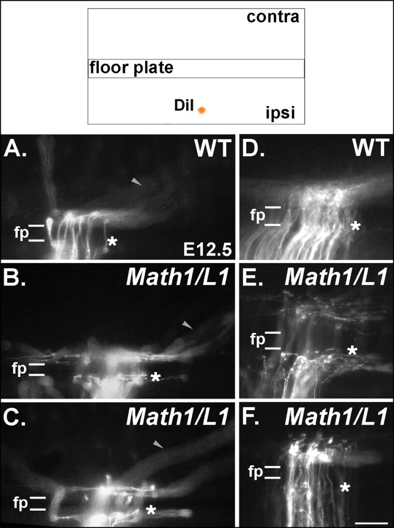
Figure 6. Mis-expression of L1 results in commissural axon projection errors in the immediate vicinity of the FP.

(Top) The pathfinding behavior of commissural axons was visualized through the application of DiI into a dorsal region of fixed, open-book spinal cord preparations taken from E12.5 embryos. (A-F) Photomicrographs of DiI-labeled axons in open book preparations derived from wild type (WT) and Math1/L1 embryos. (A, D) In WT embryos, commissural axons project directly through the FP in a well-organized fashion before turning in a predominantly rostral direction on the contralateral side of the spinal cord. (B, C, E) In Math1/L1 embryos, commissural axons “pile-up” and stall at the ipsilateral margin of the FP, or project along this boundary before ultimately crossing the FP. The arrowheads in A, B and C denote out of focus axons that are projecting along appropriately shaped contralateral trajectories in WT and Math1/L1 embryos. (F) On rare occasions, some commissural axons were observed to stall at the contralateral margin of the FP. In A-F, asterisks demarcate the position of the ipsilateral FP margin. In each panel, rostral is to the right. Scale bar in (F), 95 μm (A-C); 50 μm (D-F).
