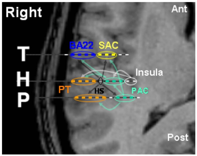
Figure 8.

Cortico-cortical connections within the auditory areas of Case 3. Arrows indicate the direction of auditory stream. Localisation of intracerebral electrodes in auditory cortex is superimposed on the patient’s MRI slices. Each dashed line (labeled T, H and P) indicates the anatomical location of a single electrode (each white segment corresponding to a given electrode contact). Each blue, yellow, red, or green line indicates which auditory region is recorded. Note that a single electrode can record cortical activity from different areas.
