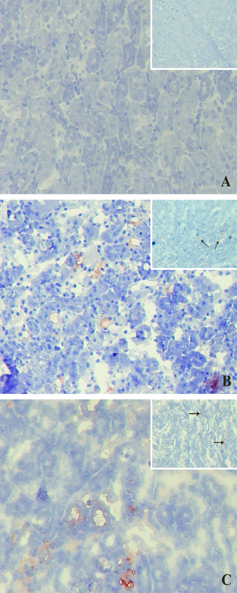
Figure 8.
MBL-C and C3 (insets) deposition in WT and Mpo−/− mice after 24 hours of reperfusion. MBL-C and C3 deposition was not different in WT (B) or Mpo−/− (C) mice. A: Virtually no MBL-C or C3 staining was detected in sham-treated WT (not shown) and Mpo−/− mice. Corresponding data were obtained for MBL-A deposition (data not shown). Shown are representative microphotographs of all groups. Original magnifications: ×200; ×100 (insets).

