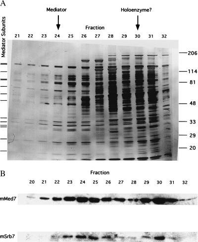
Figure 2.
Elution profiles from last step of mouse Mediator purification. (A) SDS/PAGE of TSK-Heparin-5PW fractions. Proteins were revealed by silver staining. Tick marks at left indicate positions of bands that coelute and coimmunoprecipitate (Fig. 3) and that are therefore attributed to Mediator subunits. Positions of bands due to size markers (kDa) are indicated at the right. Peak fractions of mSrb7 and mMed7 in the immunoblot (B Lower), attributed to Mediator and to possible RNA polymerase II–Mediator complex (“holoenzyme”), are indicated by arrows at the top. (B) Immunoblot of TSK-Heparin-5PW fractions with antibodies against hSrb7 and hMed7. Only the regions of the blots containing mSrb7 and mMed7 are shown.

