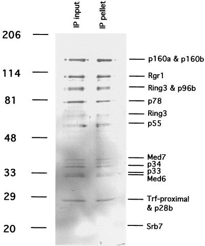
Figure 3.
Immunoprecipitation of mouse Mediator with antibodies against hSrb7. TSK-Heparin-5PW fraction 24 (50 μl) was immunoprecipitated with 100 μl of protein A-Sepharose-purified anti-hSrb7 antibodies crosslinked to 50 μl of Sepharose beads as described (5). After incubation for 4 h at 4°C in 25 mM Tris acetate, pH 7.8/200 mM potassium acetate/0.1 mM DTT/1 mM EDTA/10% glycerol/protease inhibitors, the beads were washed twice with the same buffer containing 500 mM potassium acetate/0.2% Nonidet P-40. Immunoprecipitated proteins (“IP pellet”) were eluted with 5 M urea twice for 10 min at room temperature, precipitated with trichloroacetic acid, analyzed alongside the starting fraction (“IP input,” 20 μl, precipitated with trichloroacetic acid) by electrophoresis in an 8–12% gradient SDS-polyacrylamide gel, and revealed by silver staining. Positions of bands due to size markers (kDa) are indicated at the left. Mediator subunits are identified at the right.

