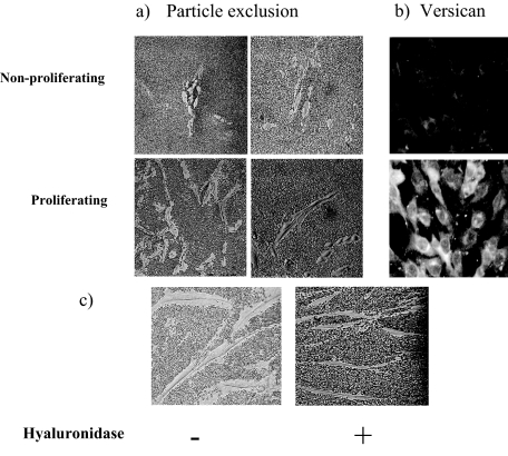
Figure 7.
Visualization of the HA pericellular coat. MCs in nonproliferating (0.2% LH) or proliferating (20% FCS) conditions were incubated with formalized erythrocytes for the particle exclusion assay (a) or incubated with anti-versican antibody and visualized with anti-rabbit-FITC IgG (b). c: Loss of pericellular coat in proliferating cells incubated with hyaluronidase as a control. Data shown are representative of five independent experiments. Original magnifications: ×200 (a); ×250 (b); ×400 (c).

