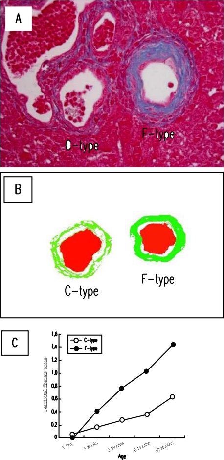
Figure 2.
Progressive fibrosis around two different type bile ducts in the PCK liver during aging. A: Periductal fibrosis around F-type bile duct was more dense than that of C-type bile duct. To determine periductal fibrosis score, the image analysis was performed as described in Materials and Methods. B: Representative analyzed image of the figure shown in A (green, fibrotic area; red, bile duct lumen). C: Fibrosis occurred progressively around both C- and F-type bile ducts during aging, in which the extent was more prominent in F-type bile duct. A, Azan-Mallory. Original magnification, ×400.

