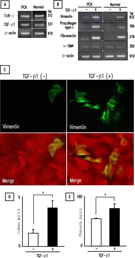
Figure 5.
Induction of mesenchymal markers in PCK cholangiocytes by TGF-β1. Cultured cholangiocytes were incubated with TGF-β1 for 3 days on type I collagen-coated cell culture dishes. A: Detection of TGF-β type II receptor (TβR-II) and TGF-β1 mRNA in PCK and normal cholangiocytes using RT-PCR. B: Effects of TGF-β1 on the expression of mesenchymal markers in cultured cholangiocytes determined by RT-PCR. C: Induction of vimentin in PCK cholangiocytes by TGF-β1. Vimentin was visualized by Alexa-488 (green), and pan-CK was visualized by Alexa-568 (red) under immunofluorescence confocal microscopy. Bottom panels were merged images of vimentin and pan-CK. D and E: Concentrations of collagen (D) and fibronectin (E) in cell culture supernatant of PCK cholangiocytes. The concentrations of collagen and fibronectin were determined as described in Materials and Methods. The data represent three independent experiments (A, B) and the mean ± SD of four per group (D, E). *P < 0.01. Original magnifications, ×1000.

