Figure 6.
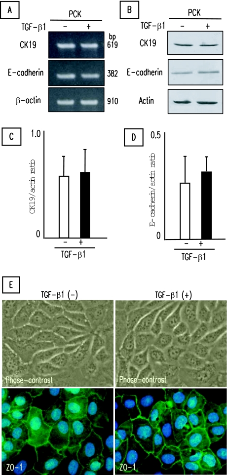
Effects of TGF-β1 on epithelial cell phenotype of PCK cholangiocytes. Cultured PCK cholangiocytes were incubated with TGF-β1 for 3 days on type I collagen-coated cell culture dishes. A and B: No inhibitory effects of TGF-β1 on the expression of CK19 and E-cadherin determined by RT-PCR (A) and Western blotting (B). C and D: Semiquantitative analysis of the results of Western blotting was performed for CK19 (C) and E-cadherin (D), confirming no significant inhibitory effects of TGF-β1. E: No effects of TGF-β1 on epithelial cell morphology of PCK cholangiocytes. E: Morphological transition was not observed after TGF-β1 treatment under the phase-contrast microscope (top). E: Tight junctions were present even between adjacent cells after the TGF-β1 treatment as shown by expression of ZO-1 (bottom). ZO-1 was visualized by Alexa-488 (green) under immunofluorescence confocal microscopy. Nuclei were stained with 4′,6-diamidino-2-phenylindole (blue). The data represent three independent experiments (A) and the mean ± SD of four per group (C, D). Original magnifications, ×1000.

