Figure 4.
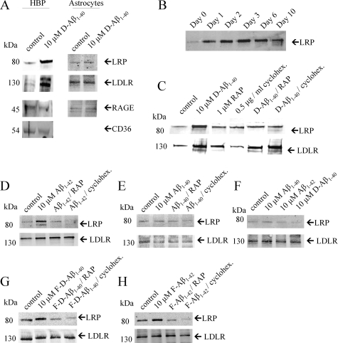
Western blot analysis of LRP-1 and LDLR expression in cultured HBPs and astrocytes. A: HBPs and astrocytes were incubated with or without 10 μmol/L D-Aβ1-40 for 3 days at 37°C. In HBPs, expression of LRP-1, LDLR, RAGE, and CD36 was observed, and both LRP-1 and LDLR were up-regulated by incubation with 10 μmol/L D-Aβ1-40. In astrocytes expression of LRP-1, LDLR, and RAGE was observed, but Aβ did not affect receptor expression. B: Treatment of HBPs with 10 μmol/L D-Aβ1-40 resulted in LRP-1 up-regulation after 1 day and sustained until 10 days after treatment with 10 μmol/L D-Aβ1-40. C: In HBPs co-incubated with RAP (1 μmol/L) or cycloheximide (0.5 μg/ml) for 3 days at 37°C, reduction of LRP-1 up-regulation was observed, whereas LDLR expression remained unaffected. D: Similar effects on both LRP-1 and LDLR expression on HBPs were observed after incubations with Aβ1-42. E: Incubation of HBPs with Aβ1-40 had no effect on both LRP-1 and LDLR expression. F: Astrocytes incubated with 10 μmol/L of Aβ1-40, Aβ1-42, or D-Aβ1-40, demonstrated no effects on both LRP-1 and LDLR expression. G and H: Incubation of either 10 μmol/L fibrillar D-Aβ1-40 (G) or Aβ1-42 (H) resulted in increased LRP-1 expression by HBPs, whereas no differences were observed for LDLR levels. In addition, co-incubation of fibrillar Aβ with RAP or cycloheximide inhibited this effect.

