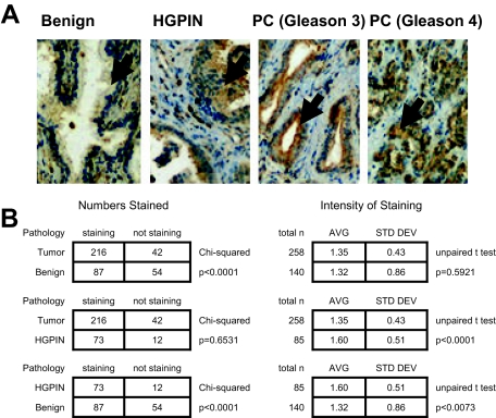
Figure 3.
Nox1 overexpression in HGPIN and cancer in human prostate tissue. A: Human prostate tissue was analyzed for Nox1 protein expression using immunohistochemical analysis as described in Materials and Methods. Arrows point to the prostate epithelial cells. Representative pictures of the normal prostate epithelial, HGPIN, and prostate cancer (Gleason 3 and Gleason 4) are shown. B: Overall NOX1 staining in benign prostate tissue, HGPIN, and prostate tumors. Analysis was performed as described in Materials and Methods. P values are a result of either χ2 or unpaired t-test analysis.

