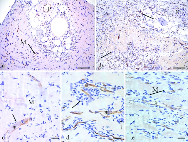
Figure 2.
CD31-labeled tissue, 2 weeks. a: Control, pedicle cross section (P) and new blood vessels (arrow) radiating into empty Matrigel (M). b: Triple growth factor treated, demonstrating a general increase in cellularity and many more new blood vessels (arrows) compared with control. P, pedicle. c: Vascular sprouts in control. Arrow indicates few accompanying cells with a new capillary (*) in relatively empty Matrigel (M). d and e: VEGF120 + FGF-2 group (d) and triple growth factor group (e), both demonstrate increased cellularity (arrows) around CD31-positive capillary sprouts (*). M, empty Matrigel. Compare with c. Scale bars: 100 μm (a, b); 20 μm (c–e).

