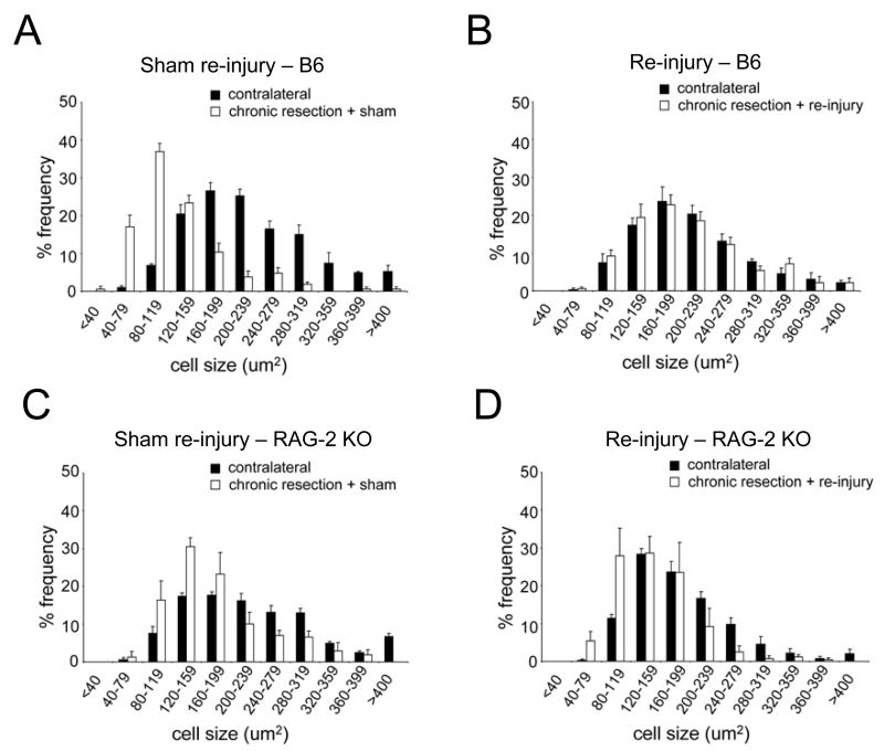
Figure 2.
Facial motor neurons binned according to cell size following sham re-injury and re-injury. Note that the distribution of neurons in the injured FMN shifts from small to large cell sizes and is normalized to the contralateral uninjured side following re-injury in B6 but not in RAG-2 KO mice.

