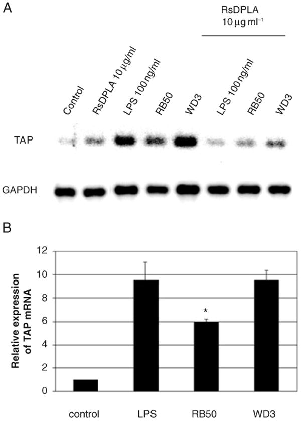
Fig. 1. Stimulation of TAP expression by LPS and bacteria via TLR4 in bovine TEC.

A. TECs were pretreated with 10 μg ml−1 RsDPLA for 1 h prior to stimulation with 100 ng ml−1 of P. aeruginosa LPS or live B. bronchiseptica for 18 h. Dose–response studies revealed that 10 μg ml−1 of RsDPLA was the lowest concentration necessary to observe the inhibition of TAP expression (not shown). Total RNA was subjected to semi-quantitative RT-PCR for 15 cycles, followed by Southern blot hybridization, as described in Experimental procedures.
B. Suppression of TAP upregulation by the type III secretion system of B. bronchiseptica. TEC were challenged with live B. bronchiseptica for 6 h. Graphical representation of the data (n = 3) indicates the mean of the fold increase over the untreated control samples ± standard error of the mean. Significance (*) of the difference between RB50 and WD3-stimulated cells was determined by t-test analysis, when P < 0.05.
