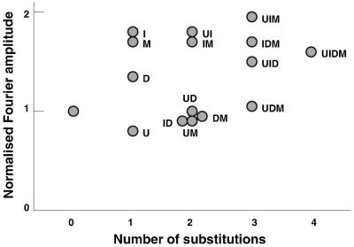
Figure 5.

Normalized Fourier amplitudes of hydroxyl-radical cleavage profiles for different substituted DNAs. The Fourier transform was calculated from densitometric profiles of the hydroxyl-radical cleavage patterns. Only the normalized amplitudes of the main harmonic are shown. The number of substitutions is specified on the abscissa as in Fig. 3. The values shown are the average of two determinations.
