
Fig. 2.
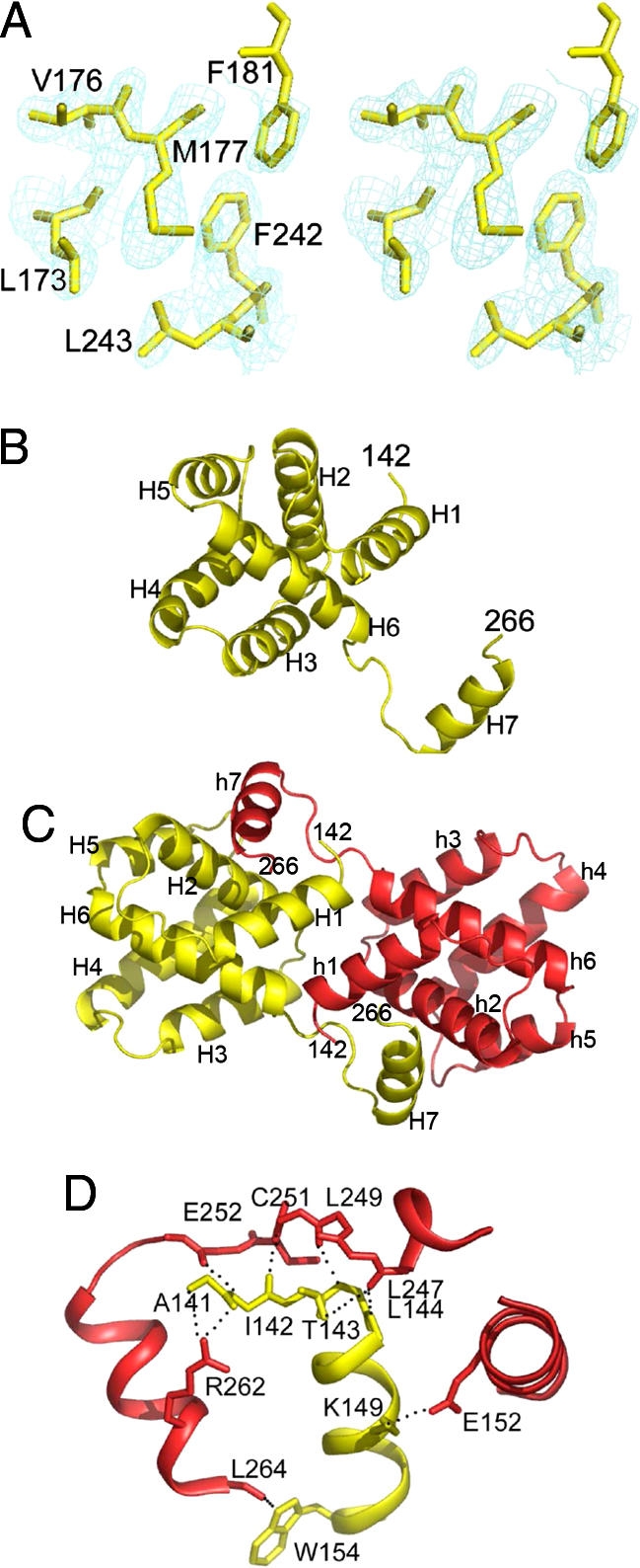
Crystal structure of dimeric VP30CTD. (A) Stereo image of the experimental electron-density map obtained from single-wavelength anomalous dispersion phases and noncrystallographic symmetry averaging showing the hydrophobic core of VP30. (B) Ribbon representation of the VP30CTD monomer that is dominated by a compact core composed of six helices and a seventh helix pointing away from the core. Secondary-structure elements are indicated. (C) Ribbon representation of the VP30CTD dimer in yellow and red. Head-to-head dimerization is supported by helix 7 interactions with the neighboring core of VP30CTD. Both N termini point away from the same side of the dimer and could potentially link up to form parallel hexamers as formed by VP3089–272 in vitro and VP30 present in EBOV particles. (D) Closeup of the VP30CTD dimerization interface. Residues mediating polar interactions are shown as all-atom model. Note that most of the interactions are between helix 7 and the loop region connecting helices 6 and 7 and the opposing helix 1.